
OUR BLOG
Valued discussions with our team from wherever you are in your day
FEATURED
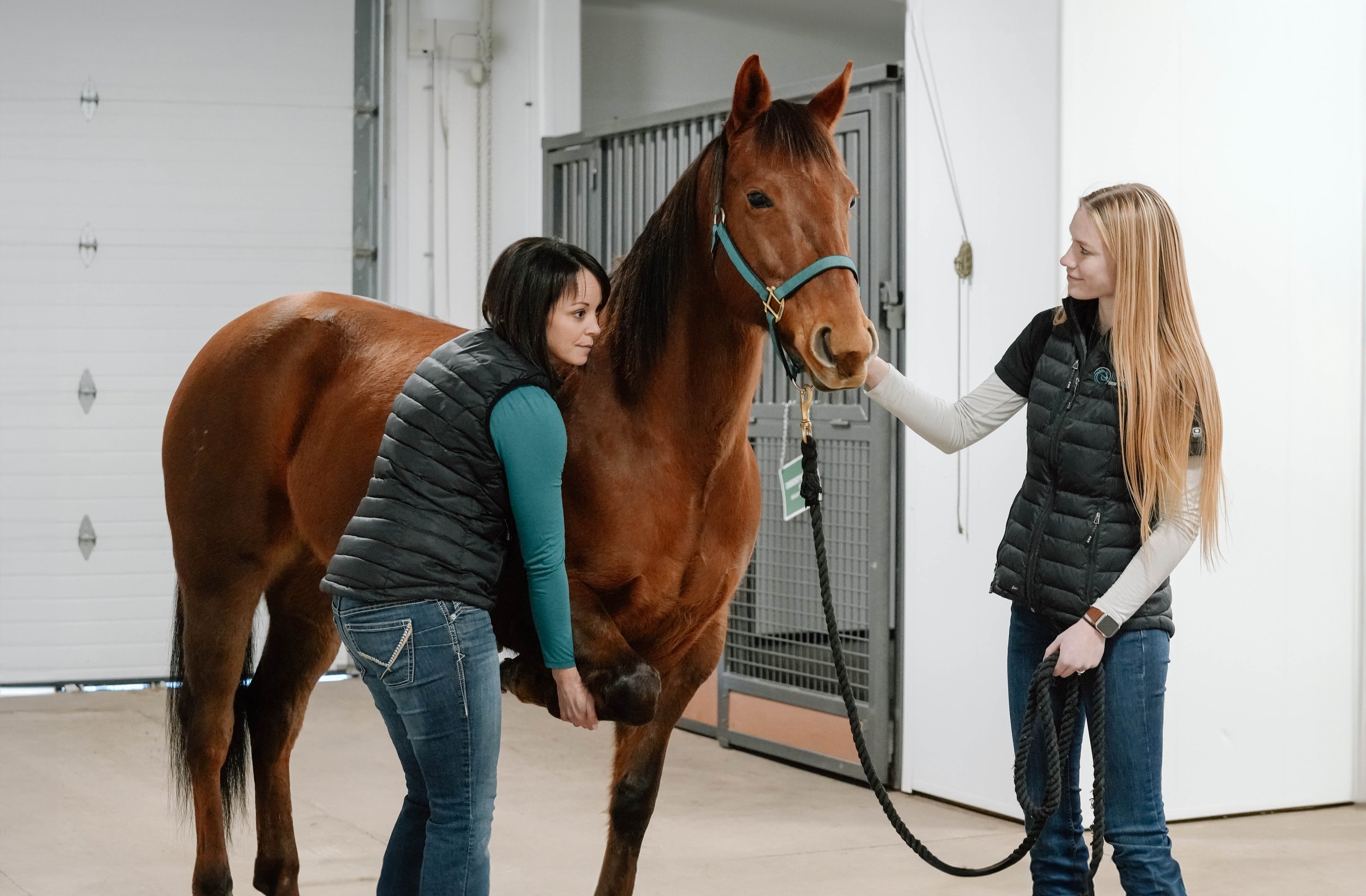
WHEN FOALING IS IMMINENT
What does a foaling actually look like? There are 3 stages to parturition:
Stage 1: this stage is the beginning of parturition where uterine contractions begin and the mare may demonstrate colic-like signs. The colic-like signs should not be violent, but include sweating, cramping, and getting up and down. These last for about 4 hours before foaling. The mare may drip and/or stream milk.
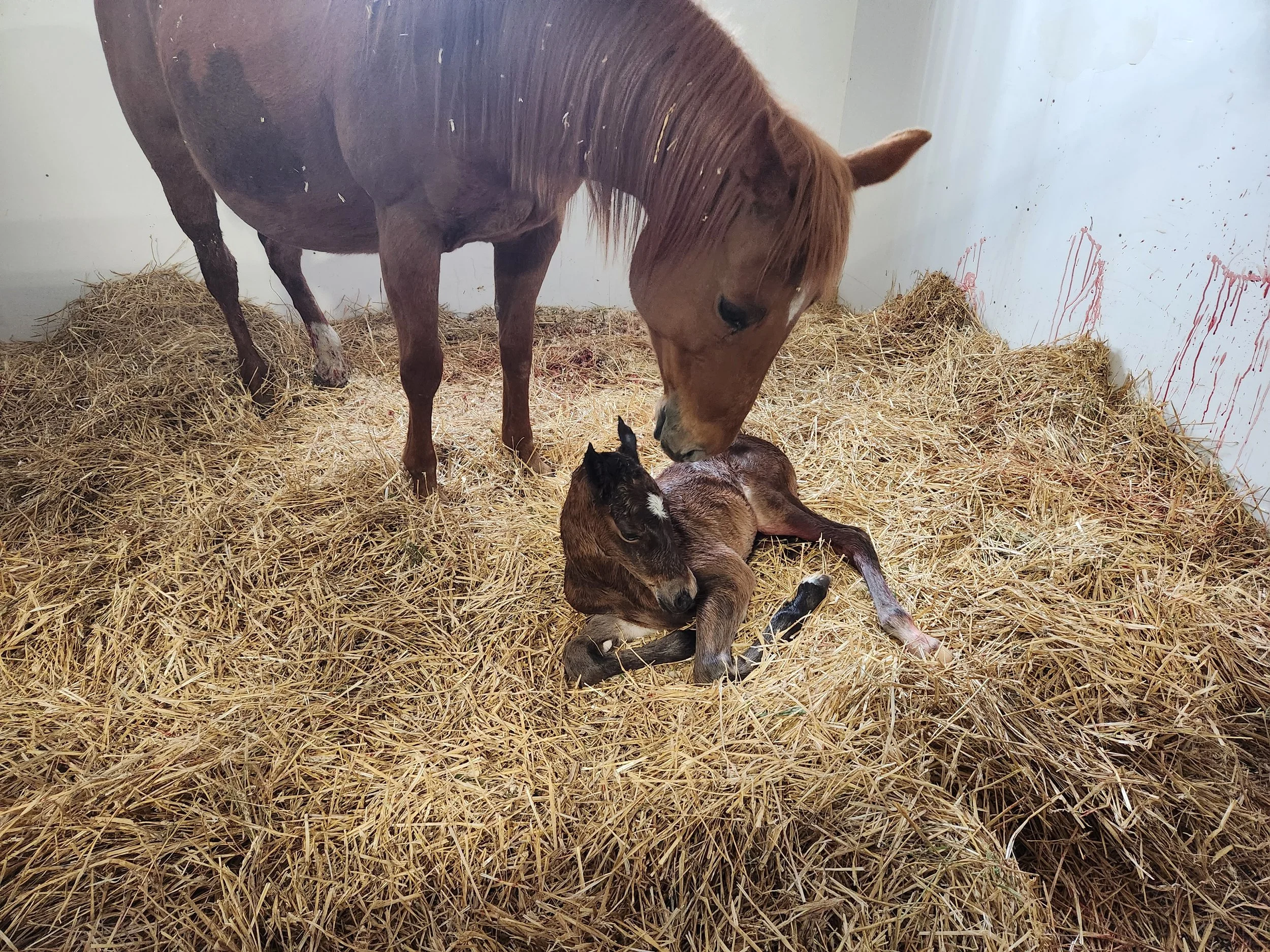
Stage 2: this stage begins with the rupture of the chorioallantios (“water breaking”) and ends once the foal is born. The foal should be born within 5-30min of the water breaking. Intervention needs to occur if there is a red bag, dystocia, or there is no progression after 30min.
Stage 3: this stage includes the passing of the placenta, or “afterbirth”. It should pass within 3 hours of foaling.


Fall Seminar 2016 - A Snapshot of the Horse's Foot - Equine Podiatry
Dr. Kirby Penttila gives an overview of equine podiatry, including a discussion of the anatomy and biomechanics of the horse's foot, the importance of shoeing survey films, and the importance of a good veterinarian-farrier-client-patient relationship. Common lameness conditions and the impact of podiatry on the treatment of these conditions are discussed in detail.

Fall Seminar 2016 - An Overview of Prepurchase Exams
Dr. Crystal Lee discusses the importance of prepurchase exams and gives an overview of what to expect in the exam.

Fall Seminar 2015 - Update on the Lameness Locator
Dr. Crystal Lee discusses scenarios in which the Lameness Locator has proven to be useful in lameness evaluation. (Fall Seminar, part 3 of 4)

Focus on Lameness - Imaging
Dr. Kirby Penttila dicusses diagnostic imaging as it relates to the lameness examination.

Focus on Lameness - Lameness Locator Demonstration
Dr. Crystal Lee performs a live demonstration of the Lameness Locator.

Focus on Lameness - The Lameness Locator
Dr. Crystal Lee discusses objective lameness evaluation using wireless inertial sensors.

Focus on Lameness - General Lameness Exam
Dr. Alyssa Butters discusses the typical progression of a lameness exam.
This excerpt is part 1 of 4

Wireless inertial sensor based objective lameness evaluation - seminar slides
We hope everyone enjoyed our Focus on Lameness Evaluation seminar on March 24, 2015! If you missed the presentation or want a second look, download the slides from the section on wireless inertial sensor based evaluation by the Equinosis Lameness Locator. Videos are coming soon!

The pre-purchase exam: A wise investment
Buying a horse can be a difficult decision. Horses seldom come with money back guarantees, so it's important to investigate before you buy. Although many horse professionals can give you an opinion on performance or breeding ability, a veterinarian will give you the best information on overall health and condition. Your veterinarian can conduct a pre-purchase examination to give you this information.
TOPIC SUBMISSIONS
Want to learn more about a specific topic? Email your ideas to our team: office@burwashequine.ca
Archive
-
Breeding
- Mar 6, 2018 Placentitis - a reason for monitoring your pregnant mare
- Mar 6, 2017 Breeding Your Mare
- Mar 20, 2015 Breeding Your Mare: A behind-the-scenes look at the science of mare reproduction
- Nov 19, 2014 Fall Seminar 2014: An Introduction to the World of Reproduction
- Nov 10, 2014 Stallion Semen Freezing
- Oct 16, 2014 Pregnant Mare Management
- Apr 24, 2014 Breeding Your Mare
-
Dentistry
- Jan 25, 2020 EOTRH: a dental disease in the elderly equine
- Nov 25, 2019 Proactive Winter Horse/Donkey/Mule Care
- Jan 22, 2017 What happens to wild horses that don't get dental care?
- Apr 25, 2014 Equine Dentistry: Why Equine Veterinarians are Uniquely Qualified
- Apr 25, 2014 A Guide To Equine Dental Care
-
Deworming
- Nov 25, 2019 Proactive Winter Horse/Donkey/Mule Care
- Feb 13, 2015 Nasty Little Parasites - An Update on Deworming
- Oct 16, 2014 Parasite Control Recommendations
-
Emergencies
- Aug 29, 2024 Equine Emergencies
- Sep 1, 2021 Getting Back to Better by Dr. Crystal Lee
- May 8, 2016 First Aid Seminar - Part 2
- May 8, 2016 First Aid Seminar - Part 1
- Apr 15, 2016 First Aid Seminar Slides
- Oct 5, 2014 Brio - Heel Bulb Laceration
- Apr 27, 2014 Stone - Septic Tarsal Sheath
- Apr 27, 2014 Luke - Puncture Wound from a Nail in the Foot
- Apr 26, 2014 Roy - Carpal Laceration
- Apr 23, 2014 Be Prepared for an Equine Health Emergency
-
Foals
- Apr 17, 2020 Fall Seminar 2019 - The Events of Normal Foaling
- Mar 6, 2018 Placentitis - a reason for monitoring your pregnant mare
- May 24, 2016 My Newborn Foal
- Apr 16, 2016 When Foaling Is Imminent
- Apr 6, 2016 When is my mare going to foal?
-
Lameness
- Mar 14, 2023 Steve with no sole
- Nov 24, 2022 Regenerative Medicine and Orthobiologics
- Jun 1, 2022 Osteoarthritis by Dr. Katy White
- Jul 1, 2020 Recognizing and Managing the Club Foot in Horses
- Jul 1, 2018 Defying Age
- Jan 23, 2016 Fall Seminar 2015 - Update on the Lameness Locator
- Sep 13, 2015 Focus on Lameness - Imaging
- Sep 13, 2015 Focus on Lameness - Lameness Locator Demonstration
- Sep 13, 2015 Focus on Lameness - The Lameness Locator
- Sep 13, 2015 Focus on Lameness - General Lameness Exam
- Apr 11, 2015 Equine Lameness Evaluation
- Mar 25, 2015 Wireless inertial sensor based objective lameness evaluation - seminar slides
-
Medicine
- Dec 27, 2024 Why Your Horse Needs Vitamin E
- Aug 29, 2024 Equine Emergencies
- Nov 24, 2022 Regenerative Medicine and Orthobiologics
- Jun 1, 2022 Osteoarthritis by Dr. Katy White
- Feb 1, 2022 Respiratory Disease Round-Up
- Nov 13, 2019 Strangles
- Dec 20, 2018 Understanding PPID
- Jul 1, 2018 Defying Age
- Feb 1, 2016 Fall Seminar 2015 - Equine Infectious Anemia
- Sep 16, 2015 Flor - Pituitary Pars Intermedia Dysfunction (PPID)
- Nov 21, 2014 Fall Seminar 2014: Pigeon Fever Updates
- Nov 4, 2014 Fall Seminar 2014: Common Conditions of the Equine Eye
-
News
- Nov 24, 2022 Regenerative Medicine and Orthobiologics
-
Podiatry
- Mar 14, 2023 Steve with no sole
- Jul 1, 2020 Recognizing and Managing the Club Foot in Horses
- Nov 12, 2016 Fall Seminar 2016 - A Snapshot of the Horse's Foot - Equine Podiatry
-
Prepurchase Exams
- Nov 5, 2016 Fall Seminar 2016 - An Overview of Prepurchase Exams
- Apr 23, 2014 The pre-purchase exam: A wise investment
-
Surgery
- May 14, 2025 Nodin - Incisor Wiring
- Sep 1, 2021 Getting Back to Better by Dr. Crystal Lee
- Sep 26, 2018 Colic Surgery
- Oct 14, 2015 Misty - Enucleation surgery
- Sep 21, 2015 Junior - Sarcoid removal
- Sep 8, 2015 Sugar - Skin Grafting
- Aug 31, 2015 Dan - Third Eyelid Removal
-
Vaccines
- Nov 25, 2019 Proactive Winter Horse/Donkey/Mule Care
- Nov 13, 2019 Strangles
- Nov 5, 2016 Fall Seminar 2016 - An Update on West Nile Virus and Rabies in Alberta
- Feb 9, 2016 Fall Seminar 2015 - Vaccines
- Apr 30, 2015 Vaccination FAQ - What are common side effects of vaccination? What can I expect after my horse is vaccinated?
- Apr 28, 2015 Why We Vaccinate - Equine Herpesvirus (“Rhinopneumonitis”)
- Apr 24, 2015 Vaccination FAQ - Why are horses vaccinated for tetanus yearly, whereas humans are boostered every 5-10 years?
- Apr 23, 2015 Vaccination FAQ - Why is the Strangles vaccine intranasal? Isn’t there an intramuscular vaccine available?
- Apr 22, 2015 Rabies in Alberta—Should We Be Vaccinating Horses?
- Apr 21, 2015 Why We Vaccinate - Equine Influenza
- Apr 19, 2015 Vaccination FAQ - Potomac Horse Fever?
- Apr 18, 2015 Vaccination FAQ - Is it better to give multiple vaccines on one date, or split them into different visits?
- Apr 17, 2015 Vaccination FAQ - My horse needs his hocks injected, and while you are here, could we vaccinate him as well?
- Apr 16, 2015 Why We Vaccinate - Strangles
- Apr 14, 2015 Vaccination FAQ - Is it okay to ride my horse immediately before or after he or she is vaccinated?
- Apr 13, 2015 Why We Vaccinate - West Nile Virus
- Apr 12, 2015 Vaccination FAQ - Can I have my horse vaccinated if (s)he has a mild “cold?”
- Apr 11, 2015 Why We Vaccinate - Eastern & Western Equine Encephalitis (“Sleeping Sickness”)
- Apr 11, 2015 Vaccination FAQ - My horse is vaccinated—why did it still get a cold?
- Apr 9, 2015 Why We Vaccinate - Tetanus in Horses
- Apr 7, 2015 Vaccination FAQ - My horse doesn’t go anywhere—does (s)he still need to be vaccinated?
- Apr 6, 2015 What's in my vaccine?
- Apr 14, 2014 Vaccination Protocols
-
Wellness
- Jun 1, 2022 Osteoarthritis by Dr. Katy White
- Feb 1, 2022 Respiratory Disease Round-Up
- Jan 18, 2022 Baled It! with Dr. Lauren Friedl
- Sep 1, 2021 Getting Back to Better by Dr. Crystal Lee
- Nov 25, 2019 Proactive Winter Horse/Donkey/Mule Care
- Dec 20, 2018 Understanding PPID
- Jul 1, 2018 Defying Age
- Feb 14, 2018 Sorting Through Supplements - how to tell if the supplement is worth buying
- Jan 8, 2017 Fall Seminar 2016 - Senior Horses - A Focus on the Care and Quality of Life of Older Horses
- Dec 8, 2016 Saying Goodbye: a discussion about euthanasia
- Jul 1, 2015 The Equine Eye by Dr. Kirby Penttila
- Apr 11, 2015 Equine Lameness Evaluation
- Oct 16, 2014 Forage Alternatives