
Dan - Third Eyelid Removal
The third eyelid, also known as the nictitating membrane, is located within the inside corner of the horse's eye. It elevates passively as the eye retracts, helping to protect the eye from trauma. There is a T-shaped piece of cartilage within the third eyelid, as well as a gland that contributes to the tear film production. Humans are in the minority here; the majority of other animal species have this third eyelid to protect their eyes.

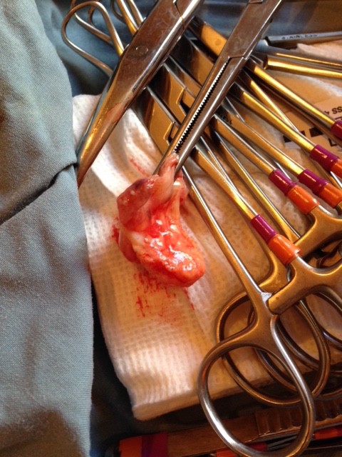
In horses, the third eyelid is prone to developing squamous cell carcinoma. Squamous cell carcinoma is the second most common tumour in horses, and it is the most common tumour in the equine eye. It develops most commonly on areas lacking pigmentation, poorly haired regions, and skin near mucocutaneous junctions. It can be quite an aggressive tumour, spreading to nearby tissues and local lymph nodes. In the third eyelid, it often initially appears as a reddened area, then becoming raised and in some cases developing a wart-like appearance. In most other areas of the body, recurrence of this tumour is extremely common unless surgical excision is combined with another treatment such as chemotherapy or cryotherapy. Fortunately, the third eyelid can be removed in its entirety, and a success rate of 90% has been reported with removal alone.
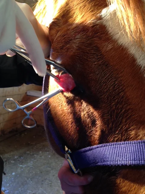
When Dan was purchased about six weeks prior to presentation, the owner noticed that the right eye would intermittently drain, and sometimes appear swollen. On examination, the majority of the eye appeared normal except for a subtle translucent area in the centre of the cornea and mild conjunctivitis (inflammation of the lining of the eyelid). The leading edge of the third eyelid had an abnormal appearance with a raised nodular component and a reddened flat component. When the signs recurred after a short course of topical antiinflammatories in the eye, the owner decided to have the third eyelid removed. At this point the presumed squamous cell carcinoma only occupied about 15% of the third eyelid, making complete surgical excision of the third eyelid a great option with an excellent prognosis.


Dan was treated at the stable under standing sedation. Topical lidocaine (local anesthesia) was applied to the cornea, and local nerve blocks were performed to numb the area. The area around the eye was scrubbed and the eye itself was lavaged with dilute betadine. The third eyelid was then grasped with forceps, locally injected with lidocaine, and clamped on either side before being transected adjacent to the clamps. The clamps were left in place for five minutes after the transection to decrease hemorrhage, and then removed.
It's very important to remove the third eyelid in its entirety, both because of the recurrence risk and because the cut edge of the third eyelid cartilage can abrade the cornea if not fully removed.

Dan was treated with oral antibiotics and antiinflammatories, as well as topical antibiotics in the right eye. There was slight hemorrhagic discharge from the eye and right nostril that resolved in a day or so.
Although there is a slight risk of increased trauma to the eye without the protection of the third eyelid, horses usually don't have a problem after surgical removal. As with many tumours, early removal in this case really increased the likelihood of success.