
Brio - Heel Bulb Laceration
Brio sustained a very deep laceration to her heel bulb while out on pasture. We examined her as soon as the owner discovered the wound, but it had likely occurred at least a week previously.

After cleaning the wound and exploring it, we were very concerned about possible involvement of the coffin joint or the digital tendon sheath. Brio was only mildly lame at the walk, which is a good sign pointing away from joint involvement given the length of time since the injury, but sometimes if a joint is open and draining the horse is not very lame because there is no pressure building up within the joint. Therefore, we distended both the coffin joint and tendon sheath with saline to check their involvement. We were able to distend both structures with fluid, without any fluid draining out of the wound, so we felt confident there was no joint involvement at that time. However, fragmented collateral cartilage could be palpated within the wound. The collateral cartilages are the large structures within the hoof (which ossify to form the ‘side bone’ we see on x-rays) shown in blue in this diagram. This finding was concerning because infection of the collateral cartilage can cause necrosis (colloquially termed quittor) and lead to chronic drainage.

Heel bulb lacerations, even without joint involvement, are a challenge to get to heal primarily because of the amount of motion in the area. Every time the horse takes a step, the wound opens and closes, preventing effective healing. Because of this, the best method of treatment is often to suture the wound and place a cast over the foot to limit movement while the wound is healing. In Brio’s case, we were unable to do this right away because of the level of infection in the wound. We first treated the wound with daily bandage changes and antibiotics for a week to allow the infection to resolve.

Once the infection was under control, we were able to surgically close the wound. We performed a nerve block for local anesthesia, and then cleaned and explored the wound and removed all pieces of fragmented collateral cartilage that we could palpate. The wound was sutured using a suture pattern specifically for wounds with a lot of tension on them, and then a cast was applied. Because Brio lives outside, it was a challenge for her owner to keep the cast dry using multiple layers of duct tape. She developed minor cast sores at the top of the cast that the owner was able to keep under control with topical treatments.


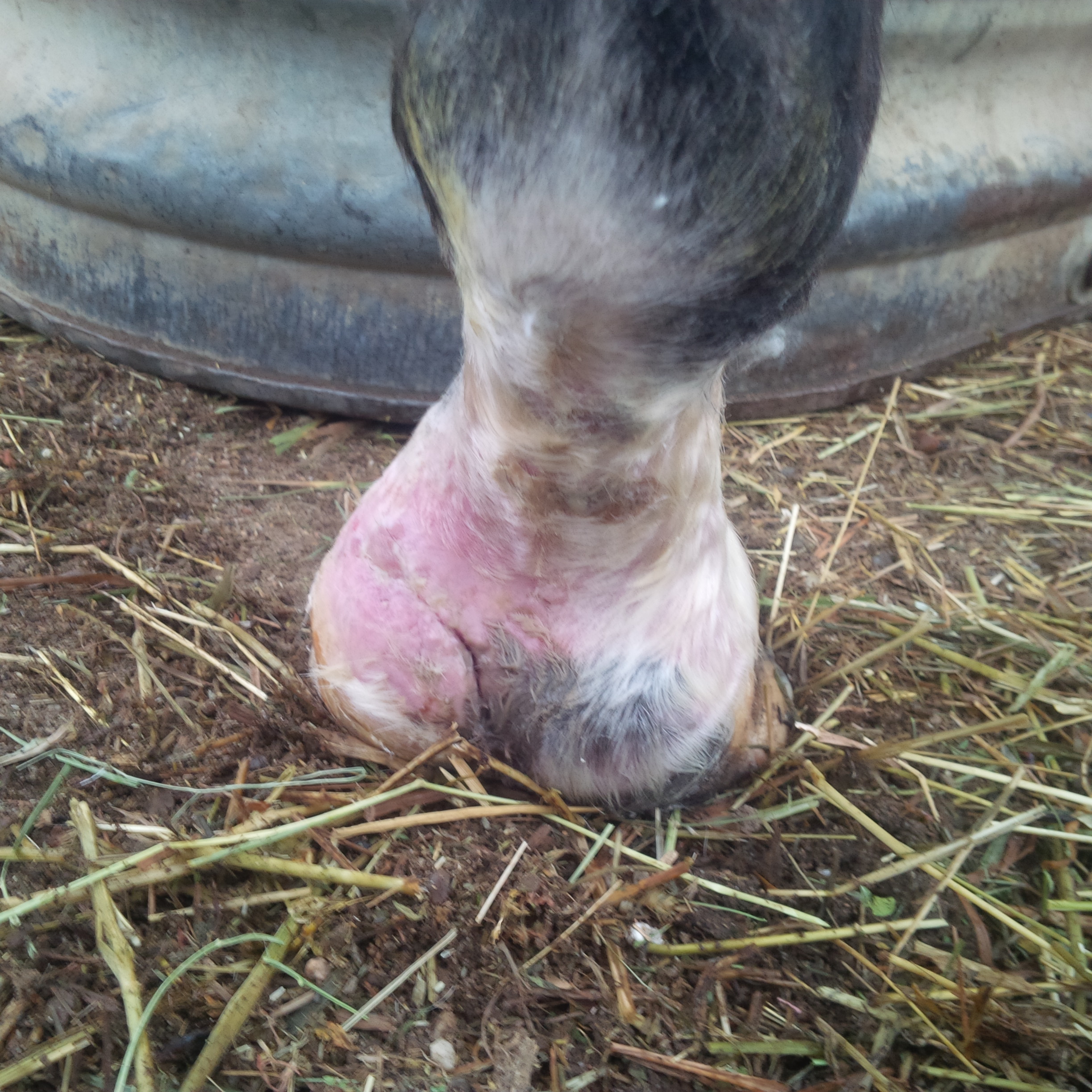
Two weeks later, the cast was removed and the sutures taken out. We were very happy to see that the wound had closed and was healing very well.
Bandages were maintained for several more weeks. We are now about four months out from the original injury, and the wound has healed very well. There has been no further drainage, which makes us much less worried about possible necrosis of the collateral cartilages. Although there will always be a scar in that area, and the hoof wall is likely to remain somewhat abnormal on that side due to the damage to the coronary band, Brio was able to return to her regular routine.
