
Luke - Puncture Wound from a Nail in the Foot
When Luke was referred to Burwash Equine by his regular veterinarian, he was unable to bear any weight on his left front limb. His veterinarian had diagnosed a puncture wound to the sole that had likely occurred several days previously. The following x-ray shows a probe placed in the puncture wound to demonstrate which structures in the foot may have been involved. Although the probe doesn’t extend the entire path of the wound, from this x-ray and taking a sample of fluid from the digital tendon sheath, we suspected infection of both the navicular bursa and the tendon sheath. (Incidentally, for those of you familiar with x-rays, Luke also has severe preexisting arthritis in both of his forelimbs that can be seen here!)
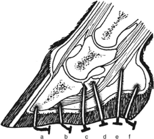
Infection of synovial structures and joints is a life threatening condition in horses. Puncture wounds to the sole can involve many different important structures of the foot (as you can see from the following image!), and should be treated as an emergency. If you find your horse with a nail in his foot, tempting though it may be to pull the nail out, the best thing to do is actually leave the nail in place and make an emergency call to your veterinarian, who can take an x-ray with the nail in place so we can determine which structures are involved before pulling the nail.
Luke underwent surgery at Burwash Equine to flush fluid through the tendon sheath and to open up a place on the bottom of the sole to allow the navicular bursa infection to drain. During this surgery we also injected fluid into the coffin joint and discovered the fluid leaking out of the puncture wound, so fluid was flushed through the coffin joint as well. Luke actually had three structures infected – his coffin joint, his navicular bursa, and his tendon sheath!
After the surgery, Luke underwent multiple days of intravenous antibiotics, regional antibiotic perfusions, flushing the structures involved, and sterile bandage changes. In the following picture, you can see the healing incision made in his sole on the left of the frog.
After almost two weeks in the clinic, Luke was walking without lameness and was able to make a triumphant return home. We were so happy to be able to take this picture! Although he still had several weeks of bandage changes to go, the infection in his foot had resolved.
