
The Case of One Thing Leading to Another...

Image 1. Radiograph of Hasty’s incsiors.
Dr. Ben Reed
Hasty Horgan is a 26 year old appendix QH gelding who recently was admitted to our clinic for extraction of three loose molar teeth that had been identified at an earlier teeth floating appointment. Exactly one year previous, Hasty had one molar tooth extracted during a routine dental exam and treatment. The loose teeth were all found to have some degree of diseased or infected tooth roots, which caused them to lose some of their attachments to the surrounding bone of the upper and lower jaw. This is commonly seen in geriatric horses whose teeth are starting to wear out. Gaps (technically called diastemas) can start to appear between teeth and provide a spot for feed to get stuck. Unless the food is removed, gum disease often develops and can result in bacteria penetrating to the root tips, causing tooth root infection and ultimately the death of all or part of the tooth. As teeth wear out, they can also develop cavities, which also allow bacteria to penetrate through to the tooth roots with similar consequences.
At both dental appointments it was noted that Hasty did not sedate particularly well and required some top ups in order to keep him sedate enough to work on. Though this can be related to an individual horse’s demeanor, Dr. Reed often feels that horses that remain somewhat awake and reactive while having a routine float performed are often experiencing some kind of oral pain. Given that the speculum that is used to keep the mouth open for floats functions by putting pressure on the incisor teeth at the front of the mouth, this response can sometimes be linked to painful incisor teeth. Interestingly, Christine, Hasty’s owner, had not noticed Hasty showing any other signs of pain, discomfort or displaying any behaviours that were unusual for him.

Images 2 and 3. Additional radiographs of Hasty’s incisors.
During the course of the molar teeth extractions, it was noted that Hasty had some abnormal changes to a few of his incisor and canine teeth. The upper incisors were showing significant age related wear with a very mild bulbous appearance to a few of the teeth at the gumline. One of Hasty’s lower canine teeth also appeared discoloured at the tip of the tooth and three of his lower incisors were very wiggly. On further examination, Dr. Reed was able to insert a 1.5” long needle directly into the pulp cavity of the canine tooth, indicating that the tooth was compromised and dead. In consultation with Christine, further radiographs were taken of Hasty’s upper and lower incisors as well as his canine teeth. These images showed that Hasty had two fractured teeth, and several more teeth that appeared abnormal, with signs of infection/deterioration, and in one case, mild enlargement. It was discussed with Christine that the radiographs indicated significant pathology in many of these teeth that could be related to early stage EOTRH (Equine Odontoclastic Tooth Resorption and Hypercemenosis), a degenerative and painful condition of incisor and canine teeth primarily seen in aged geldings (covered in a different blogpost). Alternatively, the pathologic changes in Hasty’s teeth could be due to advanced degeneration with age or due to an unknown traumatic incident in Hasty’s past. Regardless of cause, the recommendation was made, for Hasty’s comfort and health, to remove all of the affected teeth. Christine, being the conscientious and devoted horse owner that she is, consented to these further extractions. In total, six incisor teeth and one canine tooth were extracted in addition to the three molar teeth for a total of ten extractions!
Hasty tolerated the extractions extremely well and recovered from the procedure almost as if nothing had happened, displaying a strong appetite as soon as the sedation wore off.
There were a couple of interesting observations that came out of this appointment. Nerve blocks were placed in multiple locations in Hasty’s head in order to remove all sensation and pain while the extractions were being performed. While feeling for the landmarks for a nerve block to desensitize the fractured canine tooth, Hasty became very reactive just to the touch of Dr. Reed’s fingers, despite heavy sedation and other pain control meds having been given. Dr. Reed had to resort to a more distant location in order to perform a nerve block that would desensitize this tooth. For this reason, Dr. Reed believes that the canine tooth was in fact quite painful for Hasty.
Another interesting observation was made by Christine after the extractions were performed. Christine noticed that Hasty was happily eating the soaked hay he had been given in his stall at the clinic and stated that Hasty “never eats hay when he’s in a stall”. This is an observation that many owners make after the extraction of a particularly painful tooth or set of teeth, that is, the return of a more normal appetite and eating behaviour. It’s one of those things where, sometimes you don’t notice that something is wrong until after the problem has been addressed.
Though 10 extractions seems quite extreme, Hasty doesn’t seem to miss them at all! At his recheck appointment, 2 weeks following the extractions, Christine reports that she is really happy with how Hasty is doing and that he is running around in his field “as if nothing had happened”.

Images 4 and 5. Radiograph and visual of Hasty at recheck examination.
Another thing that this case is helping us to understand, is how subtle changes can sometimes indicate a much bigger problem. In many cases, it isn’t at all that the owner is negligent or unobservant, but that horses do seem to cope fairly well, even when painful conditions are present. Some theorize that as a prey animal, it is not a good thing to show the world when you are painful. Such a display may advertise that you are weakened and perhaps easy pickings. Without getting inside Hasty’s head, it is hard to know just how much those bad teeth were bugging him. But a pattern is certainly emerging of horses that are reported as “normal” by their owners, but who are found to have fairly significant pathology in some cases.
So, in summary, equine dentistry has become a much more routine part of horse care over the past few decades with newer technologies, imaging capabilities, and treatment options emerging all the time. More importantly, perhaps, is the deeper understanding that we are gaining into how a horse’s dentition factors in to their overall health and wellbeing. This post is not meant to fearmonger or shame horse owners into feeling like they need to have their horse’s teeth radiographed “just in case”, but it should help to illustrate how important regular dental exams and treatments can be for our pony pals, particularly as they age.
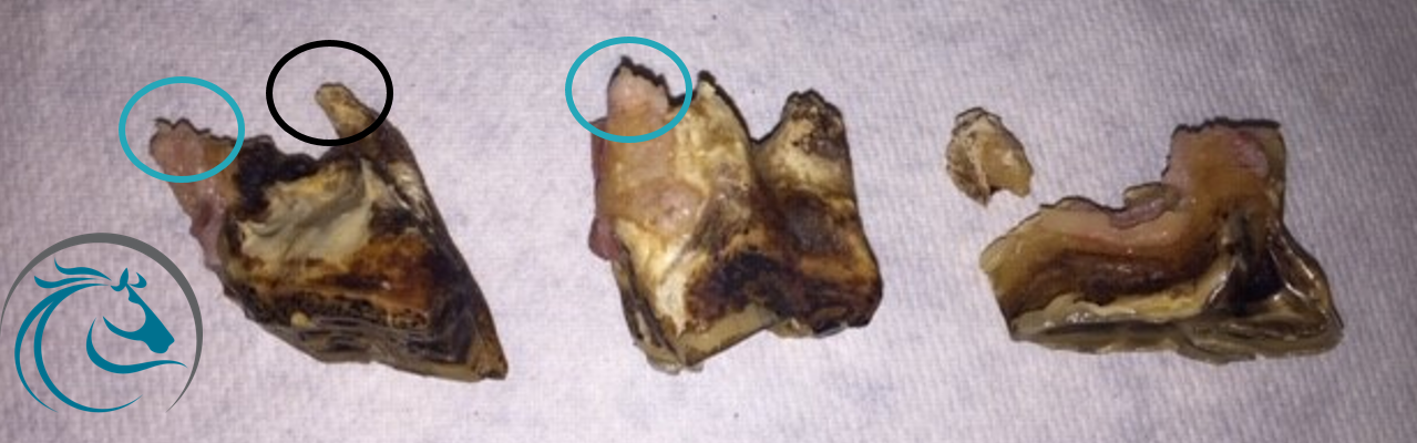
Image 6. Hasty’s extracted molar teeth. Note that some of the roots have pink tissue attached (teal circles, alive) and others do not (black circle, dead).

Images 7 (left). Several extracted incisor teeth. Image 8 (center). Note the bulbous appearance of the tooth (arrow). Image 9 (right). A fractured tooth is visible.