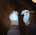
surgery

Misty - Enucleation surgery

Misty - Enucleation surgery

An enucleation is the surgical removal of a horse's eye. There are many indications for which this surgery would be performed, including trauma, neoplasia (cancer), extensive infection, or any condition causing pain in a blind eye. In Misty's case, the procedure was recommended due to an acute worsening of uveitis and ulceration along with equine recurrent uveitis flare-ups that had been occurring over the last four years.

Junior - Sarcoid removal

Junior - Sarcoid removal

Sarcoids are the most common tumour that occurs in horses. They are locally invasive, and difficult to deal with because recurrence is common even with aggressive therapy. One study showed that 14% of sarcoids occur exclusively in the periocular region (near the eye), and these tumours can be particularly tricky to deal with as it is difficult to get good margins to remove all tumour cells during surgical excision.

Sugar - Skin Grafting

Sugar - Skin Grafting

Sugar sustained a major laceration to the front of her carpus during the big snowstorm we had in September 2014. Even though her owner found it the day it happened, there was already a large amount of swelling present as well as a large amount of dirt and contamination in the wound. Initial treatment included intravenous regional limb perfusions with antibiotics, intravenous antibiotics, and bandaging. Because of the large amount of motion present on the front of the carpus, we ultimately decided to use pinch grafts in this wound. Pinch grafts are small 3mm discs of skin, harvested by removing an elevated cone of skin, that are implanted into small slits in the granulation tissue.

Dan - Third Eyelid Removal

Dan - Third Eyelid Removal

In horses, the third eyelid is prone to developing squamous cell carcinoma. Squamous cell carcinoma is the second most common tumour in horses, and it is the most common tumour in the equine eye. It develops most commonly on areas lacking pigmentation, poorly haired regions, and skin near mucocutaneous junctions. It can be quite an aggressive tumour, spreading to nearby tissues and local lymph nodes. In the third eyelid, it often initially appears as a reddened area, then becoming raised and in some cases developing a wart-like appearance. In most other areas, recurrence is extremely common unless surgical excision is combined with another treatment such as chemotherapy or cryotherapy. Fortunately, the third eyelid can be removed in its entirety, and a success rate of 90% has been reported with removal alone.

Brio - Heel Bulb Laceration

Brio - Heel Bulb Laceration

Heel bulb lacerations, even without joint involvement, are a challenge to get to heal primarily because of the amount of motion in the area. Every time the horse takes a step, the wound opens and closes, preventing effective healing. Because of this, the best method of treatment is often to suture the wound and place a cast over the foot to limit movement while the wound is healing.

Brio sustained a very deep laceration to her heel bulb while out on pasture. We examined her as soon as the owner discovered the wound, but it had likely occurred at least a week previously.

Luke - Puncture Wound from a Nail in the Foot

Luke - Puncture Wound from a Nail in the Foot

When Luke was referred to Burwash Equine by his regular veterinarian, he was unable to bear any weight on his left front limb. His veterinarian had diagnosed a puncture wound to the sole that had likely occurred several days previously. The following x-ray shows a probe placed in the puncture wound to demonstrate which structures in the foot may have been involved. Although the probe doesn’t extend the entire path of the wound, from this x-ray and taking a sample of fluid from the digital tendon sheath, we suspected infection of both the navicular bursa and the tendon sheath.