
Sarcoids are the most common tumour that occurs in horses. They are locally invasive, and difficult to deal with because recurrence is common even with aggressive therapy. One study showed that 14% of sarcoids occur exclusively in the periocular region (near the eye), and these tumours can be particularly tricky to deal with as it is difficult to get good margins to remove all tumour cells during surgical excision.
The cause of sarcoid development is multifactorial. Breed plays a role, as Quarter Horses are almost twice as likely to develop sarcoids as Thoroughbreds. Conversely, Standardbreds are less than half as likely to develop sarcoids as Thoroughbreds. As well, bovine papillomavirus has been shown to be present in nearly all sarcoid tissues. It is likely that a triggering event such as trauma is required to activate a latent viral state.

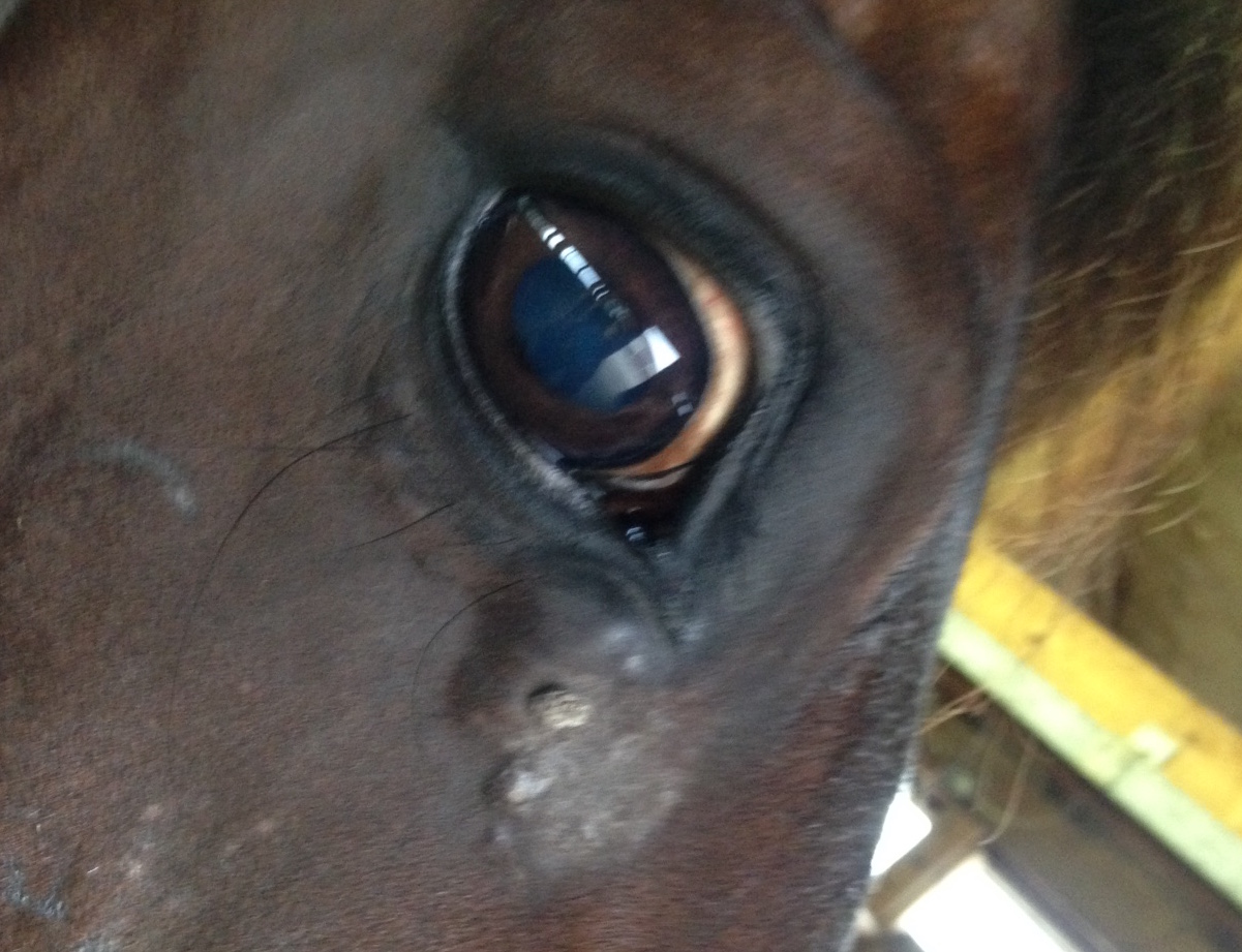
There are five broad categories of sarcoids: occult (hairless areas), verrucous (wart-like lesions), nodular fibroblastic (firm round nodules), ulcerative fibroblastic (fleshy masses that bleed easily), malevolent (the most aggressive type, which spreads quickly), and mixed (which contain more than one type). As is visible in the above photo, the sarcoid near Junior's eye was a mixed sarcoid, with nodular and verrucous components. The owner had first noticed a small wart-like lesion three months prior to presentation. It grew very quickly, and the nodular portions first appeared one month prior to presentation. The nodular portions were noticeably larger even in the three days before the exam.
Because of its location as well as the fact that the sarcoid seemed to growing so quickly, it was deemed necessary to treat it quickly and as definitively as possible. Options for treatment of sarcoids include surgical excision, cryotherapy (freezing), and topical or intralesional chemotherapy. In this case, we decided to surgically remove the tumour, and then implant cisplatin beads at intervals surrounding the incision. Since there isn't any extra tissue this close to the eye, there is a high risk of leaving tumour cells behind if surgical excision was performed without additional therapy. The cisplatin beads contain a chemotherapeutic that has been shown to provide a 75% nonrecurrence rate at two years for periocular sarcoids.

With Junior heavily sedated and standing in the stocks, nerve blocks were performed to provide local anesthesia to the area of the tumour. An incision was made around the tumour, and dissection performed around its margins. The tumour extended deeper than anticipated at the time of surgery. There was a significant amount of tension on the closure, so stents (the little pieces of tubing visible in the photo) were placed around the suture to prevent it from cutting into the skin. Three cisplatin beads were placed through tiny stab incisions above and below the main incision. These beads release chemotherapeutic medication for about six weeks after they are placed, and have not been shown to affect wound healing.
Junior was discharged the day of surgery with oral antibiotics and antiinflammatories. The sutures were removed eleven days after surgery. Due to the amount of tension on the sutures, there were sores present under some of the sutures at the time of removal, but these areas healed well with the application of antibiotic ointment. It is now just over a year after this surgery, and there is so far no sign of recurrence. It will be important for his owner to monitor carefully for any sign of recurrence of the tumour so that it can be treated immediately.
